
WHAT IS AN EOSINOPHIL ?
The eosinophil is a specialized effector cell of the immune system.
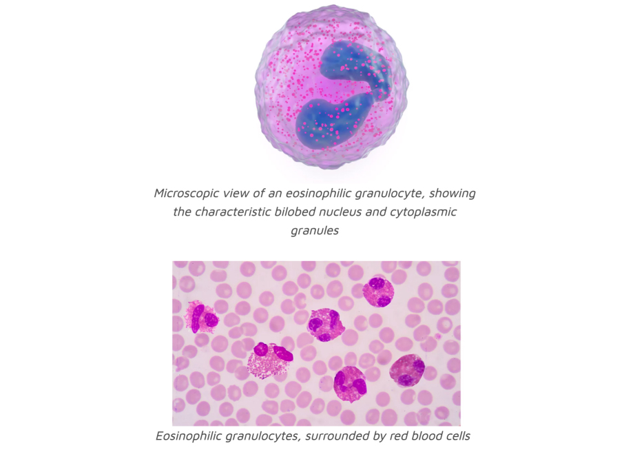
When looked at under the microscope, the eosinophil is characterized by a nucleus (contains the genetic information) with two lobes and a cytoplasm that is filled with multiple granules which contain inflammatory proteins. Activated eosinophils can degranulate and thereby release these proteins to the environment which causes tissue damage. In normal individuals, eosinophils make up about 1–3% of white blood cells.
To be seen under the microscope, blood cells are stained with hematoxylin and eosin (H&E) which is the standard coloration used in histology. The hematoxylin stains the nuclei of cells with a purple colour while the the eosin stains granular proteins in pink. The intense pink staining of the eosinophils gave them their characteristic name (eosinophil means “eosin-loving”).
Maturation of eosinophils in the bone marrow and their functions
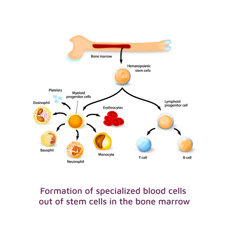
Eosinophils are formed in the bone marrow where they spend about 1 week in the process of maturation before getting access to blood vessels. Eosinophils travel through the blood vessels for 8 to 12 hours before they finally arrive at their destination tissues, where they remain for 1 to 2 weeks. The cell communication factor Interleukin 5 (IL-5) has a major role in the recruitment of eosinophils.
Eosinophils have multiple immune functions such as moving to inflamed areas, trapping substances, killing bacteria, and parasites. Furthermore, eosinophils participate in immediate allergic reactions, and they modulate inflammatory responses. Eosinophils can be either helpful (e.g. in parasite defense) or harmful (e.g. in allergic reactions) to health of an individual.
The Swiss EoE Cohort is supported by the following entities: